ㅤ
Article by Olivier Pont, Elizabeth Cordoves & Margaretha Morsink
In situ heart valve tissue engineering using a bioresorbable elastomeric implant: From material design to 12 months follow-up in sheep
Source Publication:
In situ heart valve tissue engineering using a bioresorbable elastomeric implant – From material design to 12 months follow-up in sheep, Biomaterials, 2017
Jolanda Kluin et al., Carlijn Bouten Lab
Congenital heart valve disease is a serious condition that affects millions of people worldwide, particularly infants. This disease often leads to severe complications, and sadly, it is responsible for the deaths of approximately 50,000 children each year. The current treatment options typically involve heart valve replacements, but these replacements are not ideal for children. Since they don't grow with the child’s body, patients often require multiple high-risk surgeries throughout their lives. Researchers are now exploring a new approach to solve this problem, offering hope for a better, long-lasting solution.
What did these researchers do?
The researchers in this study developed a new type of heart valve replacement that could be a game-changer. Instead of using a fully artificial valve, they designed a scaffold made of synthetic and bioresorbable materials. This scaffold is special because it serves as a mold for the patient's own cells to grow into, creating a living valve that can naturally grow and adapt as the patient does. Essentially, the scaffold helps the body regenerate its own tissue to form a valve that functions just like a healthy, natural heart valve.
Why is this important?
This research could have a huge impact on treating children with congenital heart valve disease. Traditional valve replacements do not grow with the child, meaning patients often face repeated surgeries to replace valves as they age. This creates major challenges and risks. The new approach developed by these researchers could lead to a heart valve that grows with the child, reducing the need for multiple surgeries and providing a more natural, long-lasting solution. If successful, it could dramatically improve the quality of life for children suffering from heart valve disease.
How did the researchers do this?
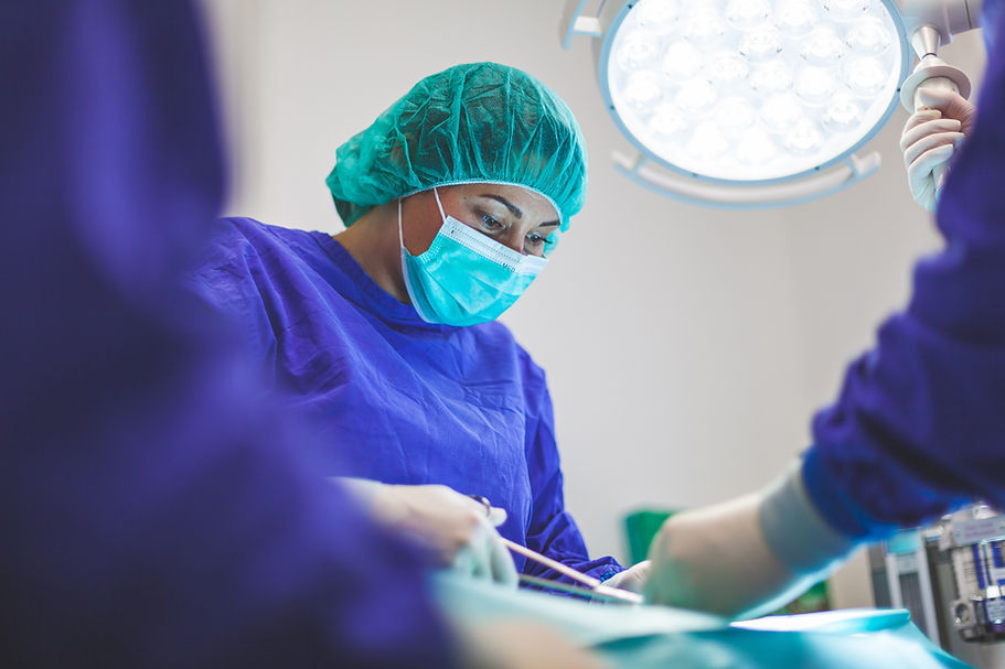
To create the new heart valve, the researchers used a scaffold made of synthetic, bioresorbable materials that break down over time. The design of the scaffold was carefully crafted to be porous, allowing the patient’s own cells to infiltrate and grow into the scaffold. This process is what helps regenerate new tissue to form a functioning heart valve. Before testing the scaffold in living animals, the scientists first tested it in the lab to evaluate its mechanical strength, biocompatibility (how well it works with the body), and degradation properties. They also simulated the conditions inside the heart using a Pulse Duplicator, a machine that mimics blood flow, to ensure the scaffold would behave properly under real-life conditions. Once the scaffold passed these tests, the researchers moved on to testing it in sheep. They implanted the scaffolds into the sheep’s hearts and followed their progress for up to 12 months to evaluate how well the scaffold supported tissue regeneration and how the valve functioned over time.

Development of a porous scaffold for heart valve regeneration that has been implanted into sheep hearts.
What comes next?
While the results from this study are promising, the researchers acknowledge that more work is needed before this technology can be used in humans. Future research will focus on confirming the long-term durability and safety of the new valve, as well as improving the scaffold materials and design. The scientists are also looking to optimize the way the tissue grows to further enhance the regeneration process. If these steps prove successful, this approach could lead to a revolutionary change in how pediatric heart valve diseases are treated, offering a more natural and less invasive solution for patients in need.